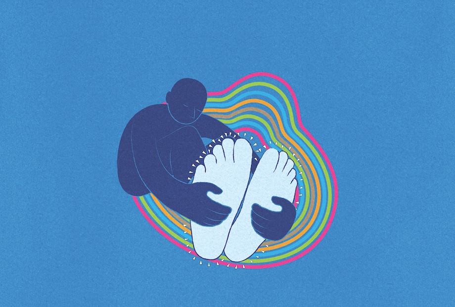
Man Experiences His Own Spine-Tingling Tale
Posted on Categories Discover Magazine

“It’s my feet,” the 50-year-old man said.
Medium height, hair tousled, Harold looked as if he had thrown on his sweatpants and loose flannel shirt in a moment of concern. Still, he appeared more intrigued than anything.
My low-key shift in the emergency department’s walk-in section had brought the usual array of back pain, ear infections, sore joints, and lacerations. This sounded no different.
“Both feet,” he continued. “I woke up this morning, and they were numb and tingly. Here.”
“Let’s step into the exam room,” I told him.
Once there, he sat down and removed his shoes.
“They don’t hurt,” he said, massaging insteps.
“Nothing else hurts? No pain anywhere else?” I asked. “No fall recently?”
“No,” he replied.
Fit and trim, Harold had no significant medical history, had walked to the hospital, and looked comfortable.
“Let’s take a look,” I suggested. Alternating between a snapped Q-tip’s sharp point and its soft cotton bulb, I probed his toes and outer feet. He accurately reported “sharps” and “dulls.” At the arches, though, he barely registered the sharp end. When I tested higher, it seemed the numbness went no farther than the ankles.
A distant alarm began buzzing in my head: Something might have happened to Harold’s spine.
The spine does more than just keep us upright and allow us to move freely — it also protects our spinal cord. That cord is comprised of nerves, the wires extending out of neurons that activate muscles or relay sensation. Nerves, in turn, are made up of bundles of axons, the individual filaments that transmit signals throughout the body. Fascinatingly, a neuron’s cell body can measure a tenth of a millimeter across, while its axons can stretch more than 3 feet in length, as in the leg’s sciatic nerve. (Imagine a starfish with arms 10,000 times longer than its body.)
Cut an axon and it might grow back; kill a neuron, however, and it’s gone forever. That’s why an injury to the spine — where neurons reside apart from the brain — can have devastating consequences.
The rest of Harold’s physical exam proved textbook normal. People get tingly feet all the time, I told myself. Tight shoes. Too much dancing. Besides the occasional backache, Harold had no history of serious spine problems. Then again, tumors can expand silently and discs can rupture for reasons as trivial as angling out of a car the wrong way. But this numbness was bilateral, and weirdly symmetrical. Did the problem begin in his feet, or somewhere up the spinal cord?
I was primed to be paranoid. Our risk-management team had recently reviewed 10 years of malpractice files and found a pattern: With missed spinal cord injuries, a recurring error had been to discount bilateral symptoms, which Harold technically had. All those patients, however, had complained of back pain and more extensive neurological deficits than a bit of foot numbness — which can be due to any number of injuries and irritations to the bones, tendons, and nerves of the feet themselves.
“When you hear hoofbeats, think horses, not zebras,” goes the adage perennially taught to med students when making a diagnosis. Common things are, well, common. And while we had identified an often-overlooked trait of an unusual beast — disc herniations that cause direct spinal cord injury —Harold’s bilateral symptoms were quite mild. Even if this was a zebra, these were pretty muffled hoofbeats.
Enough, I told myself. Harold needed an MRI of his lumbar spine.
Wear and Tear
The spine is a 30-level tower of neurons encased in a cylinder of bone. Each delicate neuronal level is encircled by a ring of bone — a vertebra — that interlocks with its mates but leaves an opening between them to the left and right. These carveouts allow the spinal cord’s neurons to send forth the nerves that provide sensation and movement to their designated body slice, or dermatome. Think of it like a centipede sprouting a pair of legs from each segment.
The chinks in our backbone’s architecture are the shock-absorbing discs. Sandwiched between the bony vertebrae, they consist of a fibrous ring and a jellylike core, or nucleus. With wear and tear, the fibrous ring can rupture, allowing the core to break into the spinal canal where the neurons live. Happily, a strong central ligament runs down the cord-facing side of the vertebrae: When a disc’s nucleus herniates, the ligament helps deflect it sideways, protecting those irreplaceable neurons in the spinal cord.
But bilateral symptoms can cause concern because they herald a disc herniating straight back into the spinal cord itself. Crush the spinal cord and all function below that level stops, which can result in paralysis of the legs and lower body, or worse. Harold’s numbness, to my alarm, suggested the straight-back scenario.
Adding to Harold’s diagnostic variables, the bony spine extends to the tailbone, but the spinal cord ends at the first lumbar vertebra, roughly at the belly button. This anatomic mismatch means that the neurons at the tip of the spinal cord bunch up, then send long nerves down to their respective bony openings — and a disc attack can theoretically happen anywhere along the way. As a result, the MRI would have to take the time to include the lower thoracic spine, around the bottom of the ribcage, as well as the lumbar spine in the lower back.
Or maybe it’s nothing, I reminded myself.
Immediate Interventions
The ER swelled with midday arrivals. Harold, third in the MRI queue, got bumped down even further by a stroke emergency that rolled in.
At one point, he grew frustrated. “I’ve been sitting here for three hours. It’s not getting worse. You sure I can’t do this as an outpatient?”
“No,” I said crisply. “If it is a disc bulging into your spinal cord, it could suddenly expand and get a lot worse. Fast.”
Harold still didn’t fit the profile of a chronic back-pain patient with rupture-prone discs. Nor could he recall recent heavy lifting or trauma. But I had already seen enough fit young patients inexplicably suffer from disc herniations.
Two hours later, the 4 p.m. shift streamed in. “It’s a long shot,” I explained to Lena, my successor. “But the numbness is very symmetrical. Please keep pushing for the MRI.”
“You bet,” she answered.
The next morning, I woke at 5:30 a.m. to check the overnight ER report. Harold’s MRI had indeed shown a disc herniation, badly compressing his spinal cord roughly around a vertebra near his waist — likely crushing his sensory neurons against the back wall of bone. The neurosurgery team wanted to operate immediately to prevent paraplegia. The problem? Harold had waited so long for the MRI and then the neurosurgeons that at 3 a.m., fed up, he had walked out of the ER.
“Oh, Christ,” I swore to myself. A few millimeters’ more disc extrusion and he’d be paralyzed. I pulled up his phone number and called, ungodly hour be damned. A raspy, exhausted voice picked up.
“Hello?”
“Harold?” I almost barked. “It’s me, Dr. Dajer. I’m really sorry about the long wait yesterday, but it’s what I feared. A herniated disc is pressing on your spinal cord. You need surgery today. Right now. Immediately. Before it gets worse.”
A pause.
“You sure?”
“Very.”
Another pause.
“I’ll call to say you’re coming. We’ll do everything possible to get you through quickly,” I urged.
That evening, Harold’s surgeons removed the offending disc. The surgery is tricky: Thoracic disc herniations are rarer than the lumbar variety — plus, ribs get in the way. The surgeons must first unroof a segment of the posterior spine to give the cord room. Then, they cut out a portion of the rib to get at the disc and extract it without harming the cord. Finally, the vertebrae above and below are braced together with screws and rods to keep the spine stable.
At some point, Harold must have wondered why the big surgery for such limited symptoms, but after three days in the hospital, he went home. For the next four weeks, he was allowed no bending or twisting. The feeling in his feet returned within days, and after three months of diligent physical therapy, the rest of him returned to normal, too.
When it comes to hoofbeats, horse or zebra, it pays to put your ear to the ground.
This story was originally published in our July August 2024 issue. Click here to subscribe to read more stories like this one.